|
How are microfossils observed?
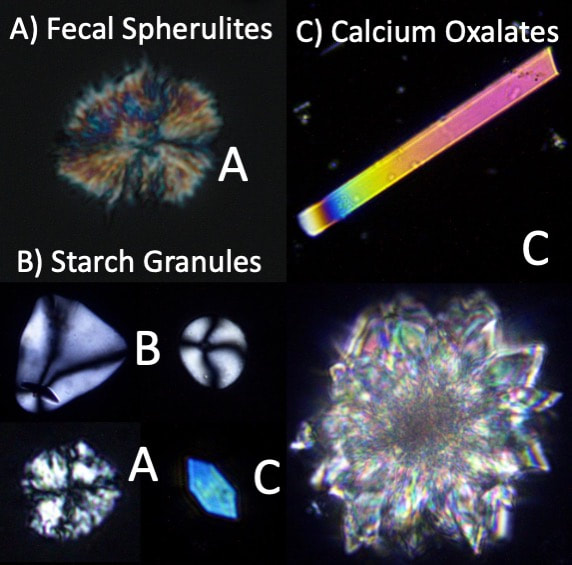
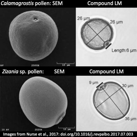
Because microfossils are so small, high-powered microscopy is necessary to observe them. Compound light microscopes consisting of an eyepiece and an objective used to view light transmitted through a microfossil are most common. Our lab typically works with compound microscopes using 200 to 500 times magnification. An essential component of a compound microscope is the ability to view objects using cross-polarized light. Microfossils like phytoliths and diatoms with amorphous (unordered) molecular structures are isotropic and go "dark" (disappear) under cross-polarized light. However, other microfossils with ordered (crystalline) molecular structures like starch granules, calcium oxalates, and fecal spherulites are anisotropic and "glow" subtly or strongly, a property called birefringence. These optical properties can be very useful in identifying microfossils as well as assessing post-depositional alteration. Some microfossils like diatoms and pollen grains have diagnostic features that are only visible at one- to several-thousand-times magnification. Observations of these types of microfossils benefit greatly from scanning electron microscopy (SEM), which uses a beam of electrons reflected off the microfossil's surface to create a highly detailed digital image. Our lab utilizes the Indiana State University, Department of Earth and Environmental Systems' SEM lab. |
